Relevance: ten years ago, artificial intelligence (AI), particularly neural networks (NN), as a diagnostic option in practice seemed a distant prospect. Today, the use of AI is becoming an increasingly popular and daily improving approach in all aspects of clinical and fundamental medicine. Purpose: design and learning of a NN to recognize four types of benign melanocytic skin tumors, integration into a mobile app to apply in practice. Material and methods: сlinical and dermatoscopic analysis of skin tumors was carried out in 600 children. In 65 cases the tumors were removed. Histological types were dermal nevus – 43% (n=28), complex nevus - 33.8% (n=22), pyogenic granuloma - 10.8% (n=7), Spitz-nevus - 6.2% (n=4), blue nevus - 3.1% (n=2), melanoma - 3.1% (n=2). Seven patients with pyogenic granulomas and two patients with melanoma were excluded. The test set included 56 dermatoscopic images. Due to the small number of images augmentation was performed. The database has been increased from 600 images to 1800. NN is written in the machine language Python. The machine learning framework was TensorFlow 2.0. The network architecture is based on the pre-trained model “EfficientNet B7”. This model uses the “supervised learning” paradigm. Each element of the sample had a class affiliation. Results: an accuracy of 83% was achieved after a period of learning on the test set. Mathematical metrics calculated in the Scikit-learn library. Sensitivity was 100% (blue nevus), 73% (complex nevus), 93% (dermal nevus), 75% (Spitz-nevus), and specificity were 98%; 94%; 82%; 98%, respectively. AI was integrated into the mobile app “KIDS NEVI”. Conclusion: AI as an auxiliary method for the skin tumors diagnosis in children and adolescents has demonstrated high potential and great opportunities. Dermatoscopic analysis of a skin tumor and a mobile app are able to provide “double control”, quick and correct clinical diagnosis and determination treatment tactics.
| Published in | European Journal of Clinical and Biomedical Sciences (Volume 10, Issue 2) |
| DOI | 10.11648/j.ejcbs.20241002.12 |
| Page(s) | 28-37 |
| Creative Commons |
This is an Open Access article, distributed under the terms of the Creative Commons Attribution 4.0 International License (http://creativecommons.org/licenses/by/4.0/), which permits unrestricted use, distribution and reproduction in any medium or format, provided the original work is properly cited. |
| Copyright |
Copyright © The Author(s), 2024. Published by Science Publishing Group |
Children, Benign Skin Tumors, Artificial Intelligence, Neural Network, Dermatoscopy
Dermatoscopic criteria | abs | % |
|---|---|---|
Typical pigment network | 5 | 7,7 |
Atypical pigment network | 3 | 4,6 |
Diameter < 2 mm | - | - |
Diameter 3-5 mm | 20 | 30,7 |
Diameter 6-8 mm | 23 | 35,4 |
Diameter 9-10 mm | 8 | 12,3 |
Diameter > 11 mm | 17 | 26,2 |
Peripheral pigment network | 2 | 3 |
Peripheral pigment globules | 7 | 10,8 |
Network break to the periphery | 1 | 1,5 |
Vascular structures in the form of comma | 22 | 33,8 |
Typical globules | 21 | 32,3 |
Atypical vascular pattern | 1 | 1,5 |
Atypical globules | 6 | 9,2 |
Homogeneous pink areas | 7 | 10,8 |
Blue-white veil | 3 | 4,6 |
Pigment blots | 16 | 24,6 |
Pigment points | 5 | 7,7 |
Asymmetry | 24 | 36,9 |
Regressive areas | 2 | 3 |
Irregular borders | 26 | 40 |
Central hyperpigmentation | 16 | 24,6 |
Irregular coloring | 51 | 78,5 |
«Cobblestone street» structure | 3 | 4,6 |
Multicolor (more than 3 colors) | 17 | 26,2 |
Homogeneous blue-gray pigmentation | 2 | 3 |
Homogeneous area of dark brown / black color | 4 | 6,2 |
Multicomponent | 10 | 15,4 |
«Starburst» | 3 | 4,6 |
Homogeneous structureless area | 33 | 50,8 |
Ulceration | 5 | 7,7 |
Histological type | Artificial intelligence | |||
|---|---|---|---|---|
precision | recall | specificity | F1-score | |
Complex nevus | 0,89 | 0,73 | 0,94 | 0,8 |
Dermal nevus | 0,84 | 0,93 | 0,82 | 0,8 |
Blue nevus | 0,67 | 1 | 0,98 | 0,8 |
Spitz-nevus | 0,75 | 0,75 | 0,98 | 0,75 |
AI | Artificial Intelligence |
NN | Neural Network |
ABCDE | A - Asymmetry, B - Border, C - Color, D - Diameter, E - Evolution |
ML | Machine Learning |
PPV | Positive Predictive Value |
TP | True Positive |
FP | False Positive |
TPR | True Positive Rate |
FN | False Negative |
TN | True Negative |
ROC curves | Receiver Operator Characteristic |
AUC | Area Under Curve |
DERM | Deep Ensemble for Recognition of Melanoma |
| [1] | Zalaudek I., Hofmann-Wellenhof R., Kittler H., Argenziano G., Ferrara G., Petrillo L., Kerl H., Soyer H. P. A Dual Concept of Nevogenesis: Theoretical Considerations Based on Dermoscopic Features of Melanocytic Nevi. J Dtsch Dermatol Ges. 2007; 5(11): 98–92. |
| [2] | Schaff er J. Update on melanocytic nevi in children. Clin Dermatol. 2015; 33(3): 368–86. |
| [3] | Haliasos H., Zalaudek I., Malvehy J., Lanschuetzer C., Hinter H., Hofmann-Wellenhof R., Braun R., Marghoob A. A. Dermoscopy of Benign and Malignant Neoplasms in the Pediatric Population. Semin Cutan Med Surg. 2010; 29(4): 218–31. |
| [4] | Kulyova S. A., Khabarova R. I. Diagnostic informativeness of the dermatoscopic pattern of skin neoplasms in children and adolescents. Russian Journal of Pediatric Hematology and Oncology. 2021; 8(4): 14–19. |
| [5] | Currie G., Elizabeth H., Rohren E. Machine Learning and Deep Learning in Medical Imaging: Intelligent Imaging. J Med Imaging Radiat Sci. 2019; 50(4): 477–487. |
| [6] | Merabishvili V. M. Malignant melanoma. Epidemiology, analytical indicators of the effectiveness of the oncological service (population study). Issues of Oncology. 2017; 63(2): 221–233. UDC 616.5-006.81: 314.4. |
| [7] | Barchuk A. A., Arseniev A. I., Belyaev A. M. et al. Efficiency of cancer screening. Issues of oncology. 2017; 63(4): 557–567. UDC 616-006.04-004.822]. |
| [8] | Ferrari A., Brecht I., Gatta G. at al. Definding and listing very rare cancers of periadric age: consensus of the Joint Action on Rare Cancers in cooperation with the European Cooperative Study Group for Pediatric rare tumors. Current Perspective. 2019; 110(1): 120–126. |
| [9] | Zukotynski K., Gaudet V., Uribe C. F. Machine Learning in Nuclear Medicine: Part 2-Neural Networks and Clinical Aspects. J Nucl Med. 2021; 62(1): 22–29. |
| [10] | Le Cun Y, Bengio Y, Hinton G. Deep Learning. Nature. 2015: 521: 436–444. |
| [11] | Barchuk A. A., Podolsky M. D., Belyaev A. M. et al. Automated diagnostics in population-based lung cancer screening. Oncology Issues, 2017; 63(2): 215–220. UDC 616.24-006. |
| [12] | Shimizu H, Nakayama KI. Artificial intelligence in oncology. Cancer Sci. 2020; 111: 1452–1460. |
| [13] | Bhinder B., Gilvary C., Madhukar N. S., Elemento O. Cancer Discov. 2021; 11(4): 900–915. |
APA Style
Khabarova, R., Kulyova, S., Senchurov, E., Mikhailova, E., Borokshinova, K., et al. (2024). Artificial Intelligence (Neural Network) in the Diagnosis of Benign Skin Tumors in Pediatric Patients. European Journal of Clinical and Biomedical Sciences, 10(2), 28-37. https://doi.org/10.11648/j.ejcbs.20241002.12
ACS Style
Khabarova, R.; Kulyova, S.; Senchurov, E.; Mikhailova, E.; Borokshinova, K., et al. Artificial Intelligence (Neural Network) in the Diagnosis of Benign Skin Tumors in Pediatric Patients. Eur. J. Clin. Biomed. Sci. 2024, 10(2), 28-37. doi: 10.11648/j.ejcbs.20241002.12
AMA Style
Khabarova R, Kulyova S, Senchurov E, Mikhailova E, Borokshinova K, et al. Artificial Intelligence (Neural Network) in the Diagnosis of Benign Skin Tumors in Pediatric Patients. Eur J Clin Biomed Sci. 2024;10(2):28-37. doi: 10.11648/j.ejcbs.20241002.12
@article{10.11648/j.ejcbs.20241002.12,
  author = {Rina Khabarova and Svetlana Kulyova and Evgeniy Senchurov and Elena Mikhailova and Kseniya Borokshinova and Alika Kulyova},
  title = {Artificial Intelligence (Neural Network) in the Diagnosis of Benign Skin Tumors in Pediatric Patients
},
  journal = {European Journal of Clinical and Biomedical Sciences},
  volume = {10},
  number = {2},
  pages = {28-37},
  doi = {10.11648/j.ejcbs.20241002.12},
  url = {https://doi.org/10.11648/j.ejcbs.20241002.12},
  eprint = {https://article.sciencepublishinggroup.com/pdf/10.11648.j.ejcbs.20241002.12},
  abstract = {Relevance: ten years ago, artificial intelligence (AI), particularly neural networks (NN), as a diagnostic option in practice seemed a distant prospect. Today, the use of AI is becoming an increasingly popular and daily improving approach in all aspects of clinical and fundamental medicine. Purpose: design and learning of a NN to recognize four types of benign melanocytic skin tumors, integration into a mobile app to apply in practice. Material and methods: сlinical and dermatoscopic analysis of skin tumors was carried out in 600 children. In 65 cases the tumors were removed. Histological types were dermal nevus – 43% (n=28), complex nevus - 33.8% (n=22), pyogenic granuloma - 10.8% (n=7), Spitz-nevus - 6.2% (n=4), blue nevus - 3.1% (n=2), melanoma - 3.1% (n=2). Seven patients with pyogenic granulomas and two patients with melanoma were excluded. The test set included 56 dermatoscopic images. Due to the small number of images augmentation was performed. The database has been increased from 600 images to 1800. NN is written in the machine language Python. The machine learning framework was TensorFlow 2.0. The network architecture is based on the pre-trained model “EfficientNet B7”. This model uses the “supervised learning” paradigm. Each element of the sample had a class affiliation. Results: an accuracy of 83% was achieved after a period of learning on the test set. Mathematical metrics calculated in the Scikit-learn library. Sensitivity was 100% (blue nevus), 73% (complex nevus), 93% (dermal nevus), 75% (Spitz-nevus), and specificity were 98%; 94%; 82%; 98%, respectively. AI was integrated into the mobile app “KIDS NEVI”. Conclusion: AI as an auxiliary method for the skin tumors diagnosis in children and adolescents has demonstrated high potential and great opportunities. Dermatoscopic analysis of a skin tumor and a mobile app are able to provide “double control”, quick and correct clinical diagnosis and determination treatment tactics.
},
 year = {2024}
}
TY  - JOUR
T1  - Artificial Intelligence (Neural Network) in the Diagnosis of Benign Skin Tumors in Pediatric Patients

AU  - Rina Khabarova
AU  - Svetlana Kulyova
AU  - Evgeniy Senchurov
AU  - Elena Mikhailova
AU  - Kseniya Borokshinova
AU  - Alika Kulyova
Y1  - 2024/09/06
PY  - 2024
N1  - https://doi.org/10.11648/j.ejcbs.20241002.12
DO  - 10.11648/j.ejcbs.20241002.12
T2  - European Journal of Clinical and Biomedical Sciences
JF  - European Journal of Clinical and Biomedical Sciences
JO  - European Journal of Clinical and Biomedical Sciences
SP  - 28
EP  - 37
PB  - Science Publishing Group
SN  - 2575-5005
UR  - https://doi.org/10.11648/j.ejcbs.20241002.12
AB  - Relevance: ten years ago, artificial intelligence (AI), particularly neural networks (NN), as a diagnostic option in practice seemed a distant prospect. Today, the use of AI is becoming an increasingly popular and daily improving approach in all aspects of clinical and fundamental medicine. Purpose: design and learning of a NN to recognize four types of benign melanocytic skin tumors, integration into a mobile app to apply in practice. Material and methods: сlinical and dermatoscopic analysis of skin tumors was carried out in 600 children. In 65 cases the tumors were removed. Histological types were dermal nevus – 43% (n=28), complex nevus - 33.8% (n=22), pyogenic granuloma - 10.8% (n=7), Spitz-nevus - 6.2% (n=4), blue nevus - 3.1% (n=2), melanoma - 3.1% (n=2). Seven patients with pyogenic granulomas and two patients with melanoma were excluded. The test set included 56 dermatoscopic images. Due to the small number of images augmentation was performed. The database has been increased from 600 images to 1800. NN is written in the machine language Python. The machine learning framework was TensorFlow 2.0. The network architecture is based on the pre-trained model “EfficientNet B7”. This model uses the “supervised learning” paradigm. Each element of the sample had a class affiliation. Results: an accuracy of 83% was achieved after a period of learning on the test set. Mathematical metrics calculated in the Scikit-learn library. Sensitivity was 100% (blue nevus), 73% (complex nevus), 93% (dermal nevus), 75% (Spitz-nevus), and specificity were 98%; 94%; 82%; 98%, respectively. AI was integrated into the mobile app “KIDS NEVI”. Conclusion: AI as an auxiliary method for the skin tumors diagnosis in children and adolescents has demonstrated high potential and great opportunities. Dermatoscopic analysis of a skin tumor and a mobile app are able to provide “double control”, quick and correct clinical diagnosis and determination treatment tactics.

VL  - 10
IS  - 2
ER  -
Children Oncology Department, N. N. Petrov National Medical Research Center of Oncology, Ministry of Health of the Russian Federation, Saint Petersburg, Russian Federation; Department of Oncology, Children’s Oncology and Radiotherapy of Saint Petersburg State Pediatric Medical University, Ministry of Health of the Russian Federation, Saint Petersburg, Russian Federation
Biography: Rina Khabarova: Cand. of Sci. (Med.), Pediatric Oncologist of the Children Oncology Department, postgraduate student of the Research Department of Innovative Therapeutic Oncology and Rehabilitation Methods of N.N. Petrov National Medical Research Center of Oncology, Ministry of Health of the Russian Federation, Assistant of the Department of Oncology, Children’s Oncology and Radiotherapy of Saint Petersburg State Pediatric Medical University.
Children Oncology Department, N. N. Petrov National Medical Research Center of Oncology, Ministry of Health of the Russian Federation, Saint Petersburg, Russian Federation; Department of Oncology, Children’s Oncology and Radiotherapy of Saint Petersburg State Pediatric Medical University, Ministry of Health of the Russian Federation, Saint Petersburg, Russian Federation
Biography: Svetlana Kulyova: Dr. of Sci. (Med.), Associate Professor, Head of Pediatric Oncology Department, Leading Researcher of the Research Department of Innovative Therapeutic Oncology and Rehabilitation Methods, Professor of the Training and Methodology Department of N.N. Petrov National Medical Research Center of Oncology, Head of the Department of Oncology, Children’s Oncology and Radiotherapy of Saint Petersburg State Pediatric Medical University, Chief Freelance Children's Specialist Oncologist of Saint-Petersburg Health Committee.
Children Oncology Department, N. N. Petrov National Medical Research Center of Oncology, Ministry of Health of the Russian Federation, Saint Petersburg, Russian Federation; Department of Oncology, Children’s Oncology and Radiotherapy of Saint Petersburg State Pediatric Medical University, Ministry of Health of the Russian Federation, Saint Petersburg, Russian Federation
Biography: Evgeniy Senchurov: Pediatric Oncologist of the Children Oncology Department, postgraduate student of the Research Department of Innovative Therapeutic Oncology and Rehabilitation Methods of N.N. Petrov National Medical Research Center of Oncology, Ministry of Health of the Russian Federation, Assistant of the Department of Oncology, Children’s Oncology and Radiotherapy of Saint Petersburg State Pediatric Medical University.
Children Oncology Department, N. N. Petrov National Medical Research Center of Oncology, Ministry of Health of the Russian Federation, Saint Petersburg, Russian Federation
Biography: Elena Mikhailova: Pediatric Oncologist of the Children Oncology Department, postgraduate student of the Research Department of Innovative Therapeutic Oncology and Rehabilitation Methods of N.N. Petrov National Medical Research Center of Oncology, Ministry of Health of the Russian Federation.
Children Oncology Department, N. N. Petrov National Medical Research Center of Oncology, Ministry of Health of the Russian Federation, Saint Petersburg, Russian Federation
Biography: Kseniya Borokshinova: Pediatric Oncologist of the Children Oncology Department, postgraduate student of the Research Department of Innovative Therapeutic Oncology and Rehabilitation Methods of N.N. Petrov National Medical Research Center of Oncology, Ministry of Health of the Russian Federation.
Children Oncology Department, N. N. Petrov National Medical Research Center of Oncology, Ministry of Health of the Russian Federation, Saint Petersburg, Russian Federation
Biography: Alika Kulyova: Postgraduate Student of the N.N. Petrov National Medical Research Center of Oncology, Ministry of Health of the Russian Federation.
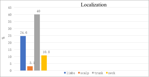
Figure 1. Distribution of tumor localization (16 (24.6%) on the limbs, 61.5 (40%) on the trunk, 2 (3.1%) on the scalp, and 7 (10.8%) on the neck).
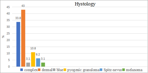
Figure 2. Distribution of histological type (dermal nevus was detected in 43% of cases (n = 28), complex nevus in 33.8% (n = 22), pyogenic granuloma in 10.8% (n = 7), Spitz-nevus in 6.2% (n = 4), blue nevus in 3.1% (n = 2), melanoma in 3.1% (n = 2).
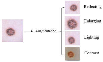
Figure 3. Augmentation.
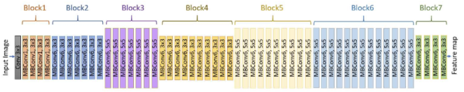
Figure 4. The architecture model “EfficientNet B7”.
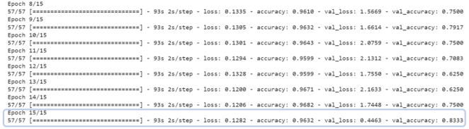
Figure 5. Adjustment of synaptic weights, 83% accuracy on the test set.
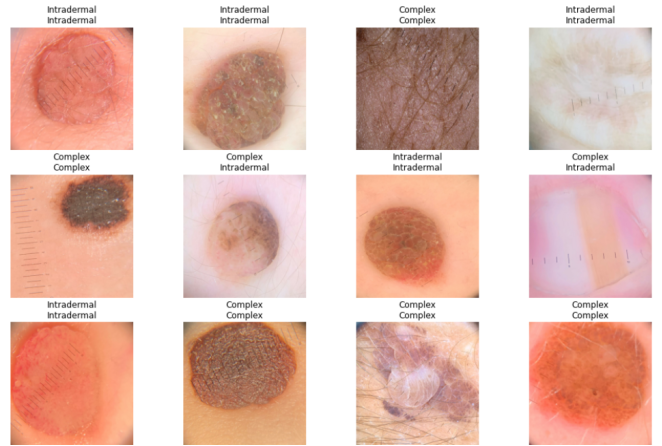
Figure 6. Neural network model after learning.
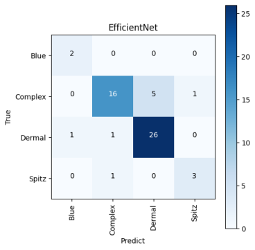
Figure 7. Confusion matrix.
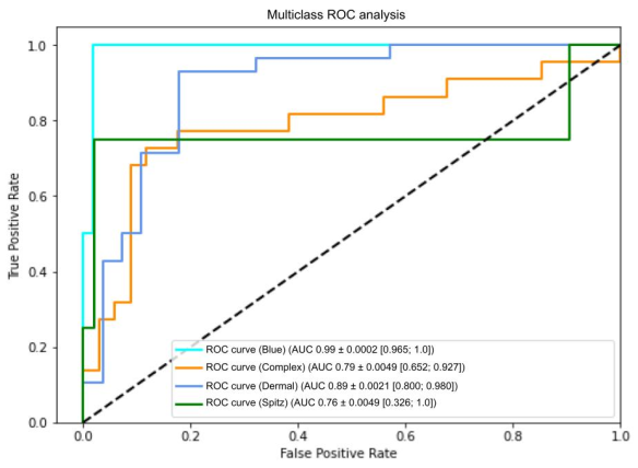
Figure 8. ROC curve, AUC. *AUC - area under the curve, (blue nevus - 0.99; complex nevus - 0.79; dermal nevus - 0.89; Spitz-nevus - 0.76).
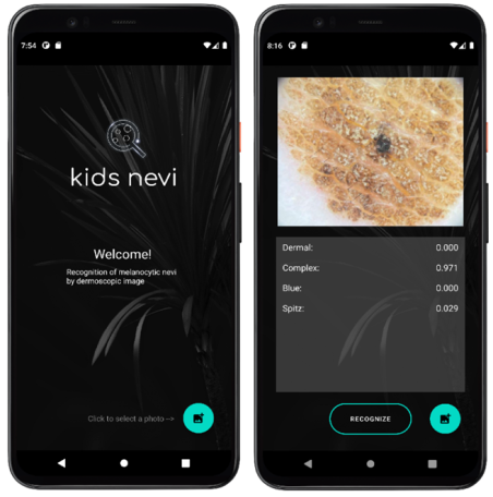
Figure 9. Mobile app “Kids nevi”.